Hernia Surgeries – (Laparoscopic/Open)

Hernia Surgeries – (Laparoscopic/Open)

A hernia happens when an internal organ or tissue pushes through a weak spot in the muscle or connective tissue that normally holds it in place. This causes a bulge or lump that may be visible or palpable. Hernias can occur in different parts of the body, and their symptoms, causes, and treatments vary depending on the type.

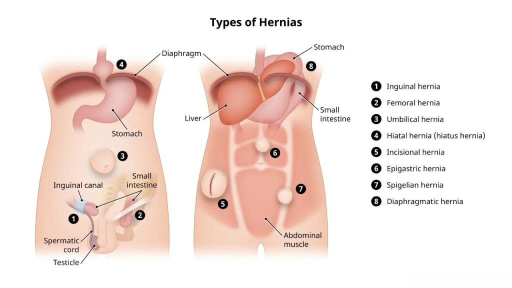
Common Types of Hernias


• Inguinal Hernia: The most common type, especially in men, appears when abdominal tissue pushes through the groin’s lower abdominal wall. Symptoms include a groin bulge and discomfort during movement or lifting.

• Ventral Hernia: Occurs anywhere on the anterior abdominal wall (front belly area), often at previous surgical sites or natural weak points. It causes bulging and sometimes pain.

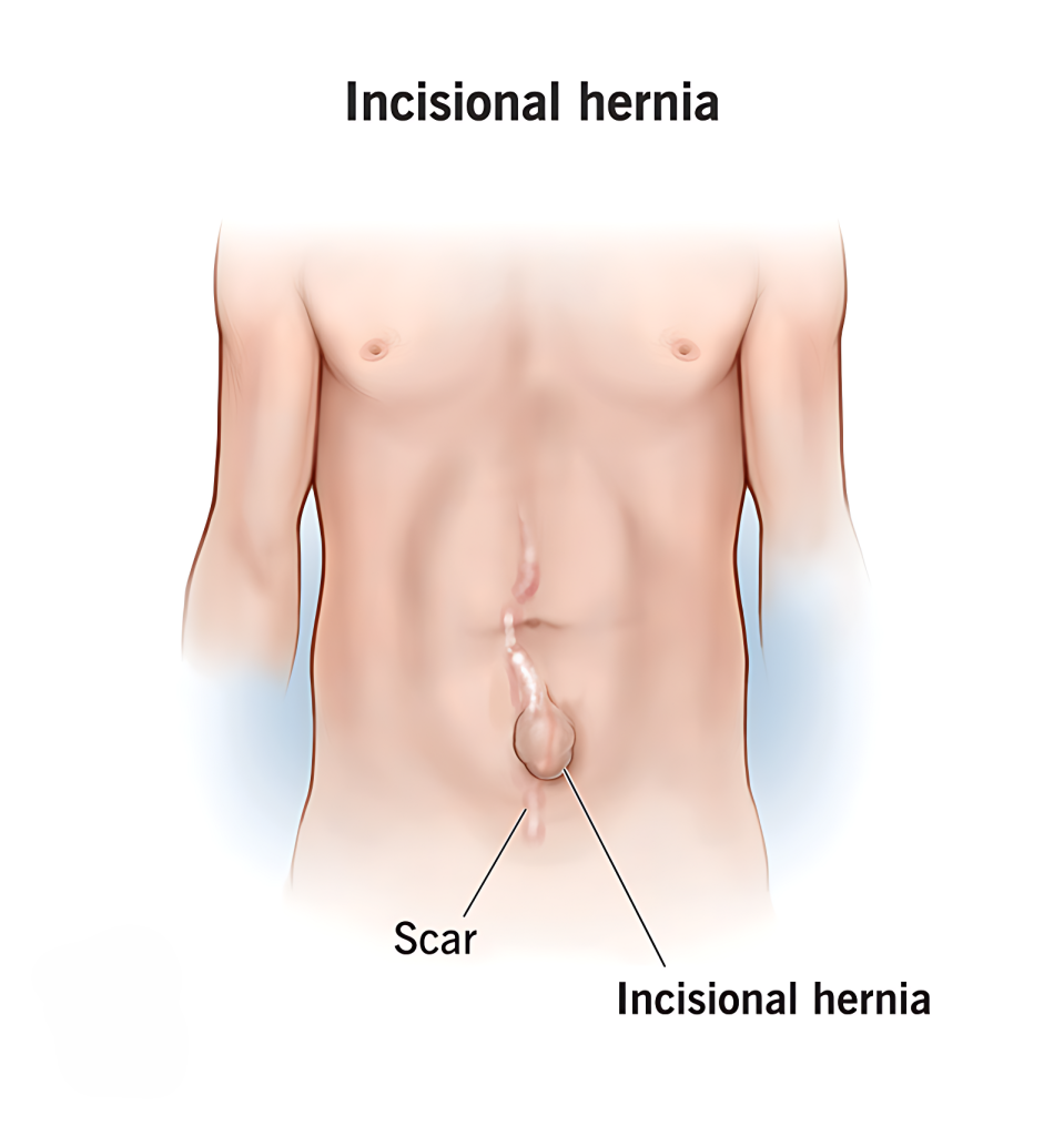
• Incisional Hernia: Develops through weakened tissue at the site of a prior surgical incision, resulting in a lump near the scar and discomfort.

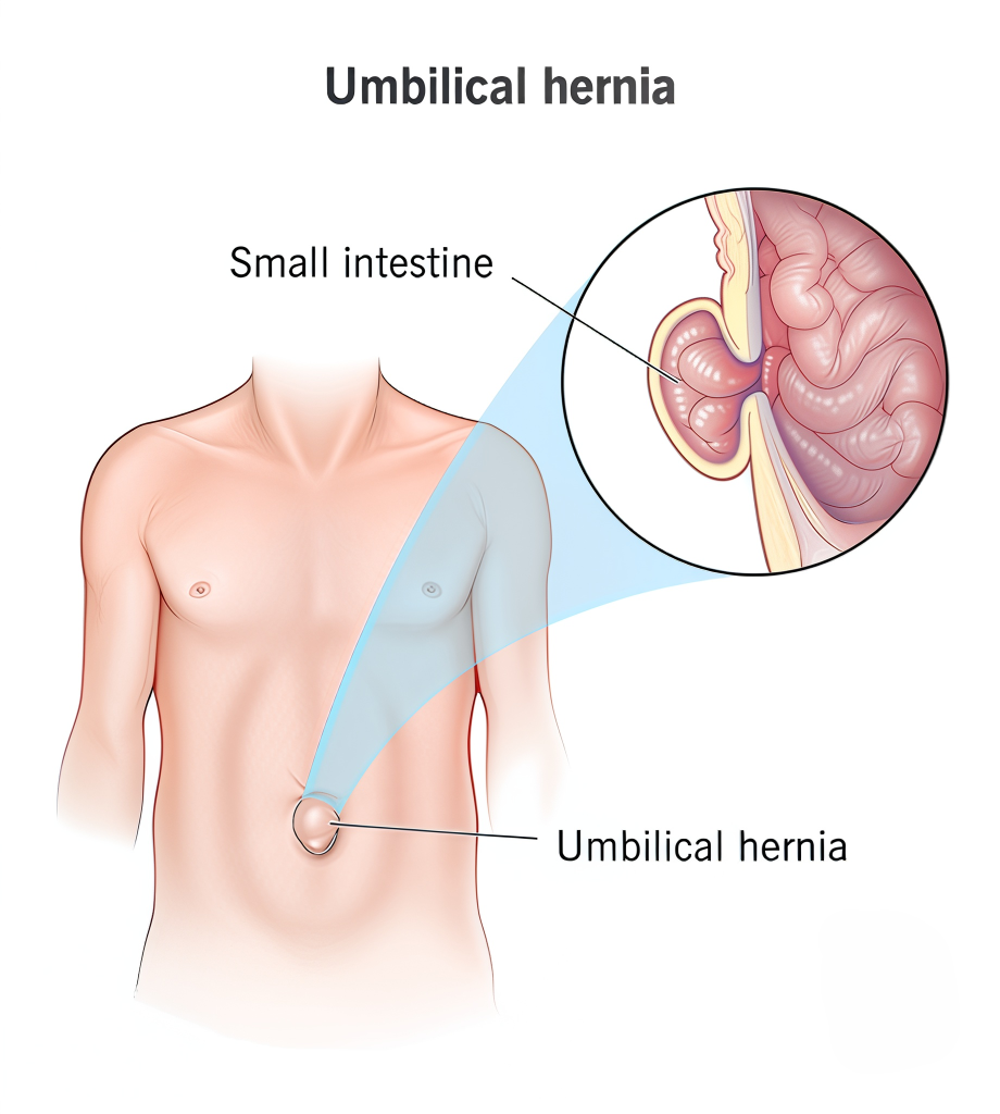
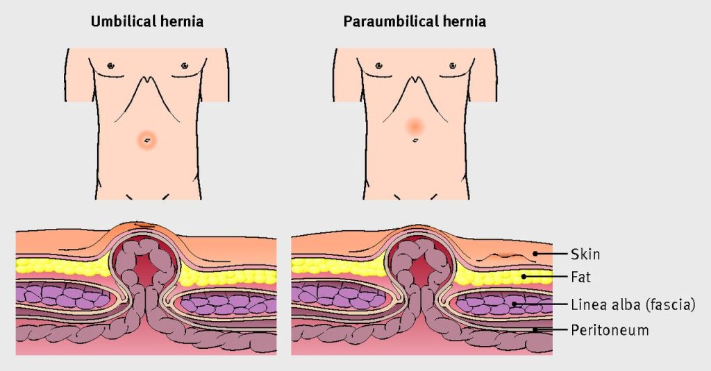
• Umbilical Hernia: Occurs near the belly button when abdominal tissue protrudes through the muscle weakness around the navel. Common in infants and adults with increased abdominal pressure.

• Paraumbilical Hernia: Similar to umbilical hernia but occurs adjacent to the navel, usually in adults.

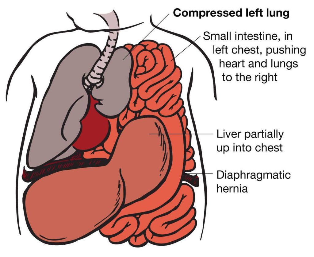
• Diaphragmatic Hernia: Abdominal organs push through a defect in the diaphragm, affecting breathing and causing chest symptoms. Can be congenital or acquired.

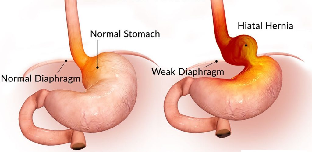
• Hiatal Hernia: Part of the stomach moves up through the diaphragm into the chest cavity, often causing acid reflux and heartburn.

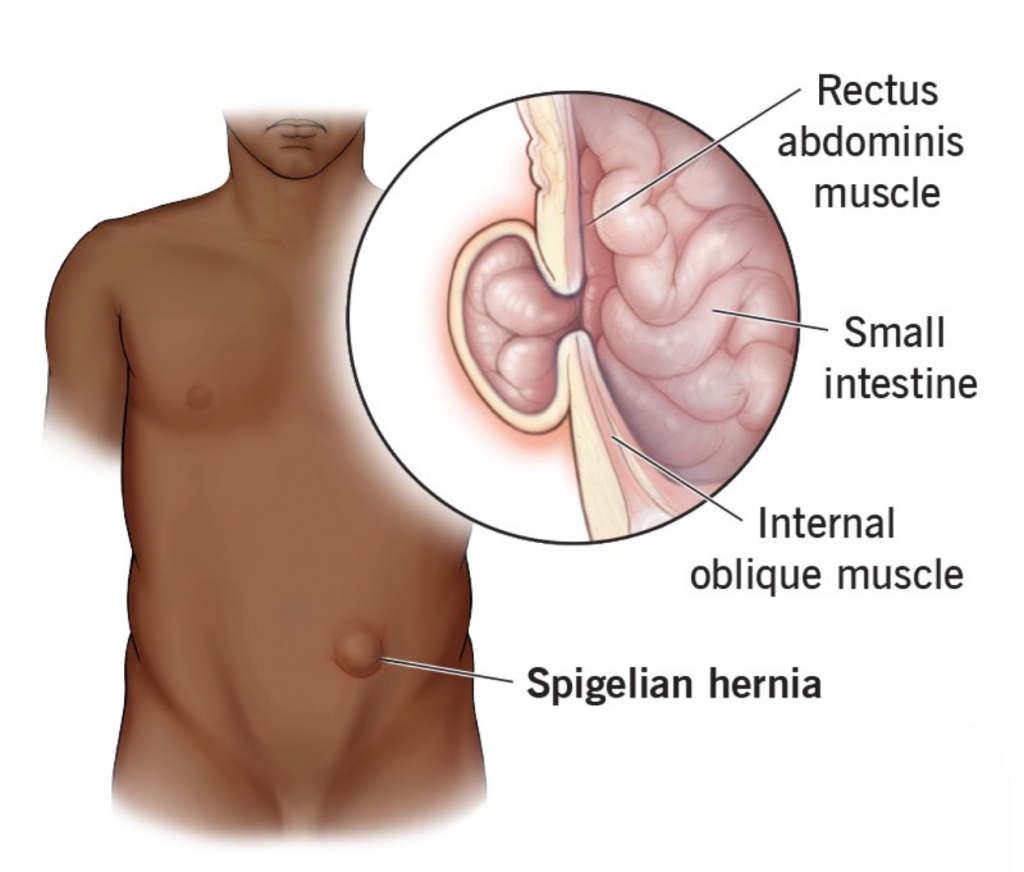
• Spigelian Hernia: A rare hernia along the abdominal muscles’ side (Spigelian fascia), difficult to detect by touch, and may cause localized pain.

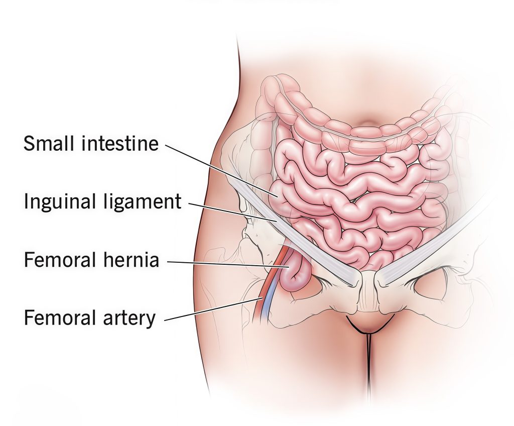
• Femoral Hernia: Tissue pushes through the femoral canal near the upper thigh, more common in women and prone to complications like strangulation.
WhatsApp Image 2025 10 01 at 5.19.30 PM

Why and When to See a Surgeon

Hernias typically do not heal on their own and may worsen over time. It is important to seek medical attention:

• If you notice a bulge or lump in your abdomen or groin.
• When experiencing pain, discomfort, or a dragging sensation around a bulge.
• If you have sudden sharp pain, tenderness, redness, nausea, or vomiting – signs of a strangulated or incarcerated hernia which is a medical emergency requiring immediate surgery.
• When symptoms interfere with daily activities or cause significant discomfort.

Early surgical consultation allows for timely repair, preventing complications like tissue strangulation or bowel obstruction. Surgery options include open repairs or minimally invasive laparoscopic procedures, both aiming to return protruding tissue safely and strengthen the abdominal wall.
Inguinal hernia 1
An inguinal hernia occurs when tissue, such as part of the intestine or fatty tissue, pushes through a weakened spot in the lower abdominal muscles near the groin area. This causes a noticeable bulge, often more visible when standing, coughing, or straining. Inguinal hernias are one of the most common types of hernias, especially affecting men due to the natural anatomy of the groin region.

The hernia forms where the abdominal wall is weaker, either due to a congenital defect present at birth or a weakness acquired over time because of strain on the muscles. Common risk factors include heavy lifting, chronic coughing, obesity, aging, and previous abdominal surgeries. In men, the hernia may extend into the scrotum, causing pain and swelling.

Symptoms of an inguinal hernia include a groin bulge accompanied by discomfort, a heavy or dragging sensation, and occasional pain that worsens during physical activities like bending or lifting. Sometimes, the hernia may cause a burning or aching feeling in the groin area. In more severe cases, if the hernia becomes trapped or strangulated, it can lead to sudden pain, nausea, vomiting, and requires immediate medical attention.

Diagnosis is primarily through clinical examination and sometimes supported by imaging tests. Treatment usually involves surgical repair to push the herniated tissue back and strengthen the abdominal wall, often using a mesh to prevent recurrence. Early treatment is important to avoid complications and improve quality of life.
Ventral hernia
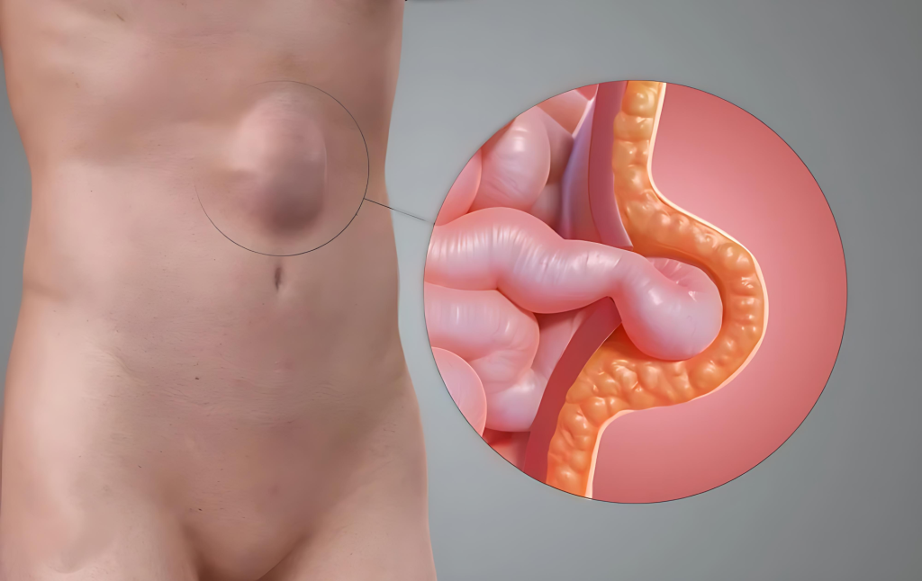
A ventral hernia occurs when tissue or part of an internal organ pushes through a weakness or opening in the muscles of the front abdominal wall. This results in a visible bulge or lump in the belly area, which may be soft or tender to touch. The term “ventral” refers to the front side of the body, meaning this type of hernia can appear anywhere along the abdominal wall.

Ventral hernias can develop as a result of a natural weakness present from birth or due to factors that weaken the abdominal muscles over time. Common causes include previous abdominal surgeries (leading to incisional hernias), pregnancy, obesity, injury, or persistent straining from heavy lifting or chronic coughing.

Symptoms vary but often include a noticeable bulge in the abdomen that may grow larger with activity or strain. Some people may experience discomfort, pain, or a feeling of pressure around the site of the hernia. In serious cases, the hernia can become trapped or strangulated, leading to sharp pain, nausea, vomiting, and requiring immediate medical attention.

Diagnosis typically involves a physical examination and sometimes imaging tests like an ultrasound or CT scan. Treatment usually requires surgical repair to close the defect in the abdominal wall, often reinforced with a mesh to reduce recurrence. Surgery can be done through a traditional open approach or minimally invasive laparoscopically. Early diagnosis and treatment are important to relieve symptoms, prevent complications, and restore strength to the abdominal wall for a return to normal activities.
incisional hernia
An incisional hernia is a type of hernia that develops at or near the site of a previous surgical incision in the abdominal wall. After surgery, the muscles and tissues around the incision need time to heal and regain strength. However, if this healing is incomplete or weak, it can allow internal tissues or organs, such as parts of the intestine or fatty tissue, to push through the weakened area, forming a bulge or lump.

This condition is a relatively common complication following abdominal surgeries, especially larger or emergency operations. Incisional hernias may appear weeks, months, or even years after the initial surgery, often becoming more noticeable when standing, coughing, or engaging in physical strain.

Common symptoms of an incisional hernia include a visible bulge near the surgical scar, discomfort or aching around the area, and sometimes sharp pain during movement or heavy lifting. In some cases, more serious complications can occur if the hernia becomes trapped, causing severe pain, nausea, and requiring urgent medical intervention.

Several factors can increase the risk of developing an incisional hernia, such as obesity, smoking, diabetes, infections at the surgical site, or premature physical activity before complete healing. Proper care after surgery including rest, avoiding heavy lifting, and managing chronic health conditions can help reduce this risk.

Treatment usually involves surgical repair to close the defect in the abdominal wall and reinforce it, often using a mesh for added strength. Surgical techniques may vary from open repair to minimally invasive laparoscopic procedures depending on the hernia’s size, location, and the patient’s overall health. Early diagnosis and appropriate treatment are essential to prevent complications and restore abdominal strength, helping patients return to daily activities safely.
umbilical hernia
An umbilical hernia is a condition where a part of the intestine or fatty tissue pushes through a weak spot in the abdominal muscles near the belly button (navel). This causes a soft bulge or swelling around the navel area, which may become more obvious when coughing, laughing, or straining. Umbilical hernias are very common, especially in infants, but they can also occur in adults.

In infants, the hernia happens because the muscles around the umbilical cord opening do not close completely after birth. Most infant umbilical hernias close on their own by the age of two, and they typically cause no pain. However, in adults, an umbilical hernia usually develops due to increased pressure on a weak spot in the abdominal wall, which can be caused by factors like obesity, multiple pregnancies, heavy lifting, or persistent coughing.

Symptoms in adults may include a noticeable bulge near the belly button, discomfort, pain, or a feeling of pressure. While most umbilical hernias are painless, complications such as incarceration or strangulation (where the herniated tissue becomes trapped or loses blood supply) can cause severe pain and require urgent treatment.

Diagnosis is made through a physical examination, and imaging tests may be used if the diagnosis is unclear or to check for complications. Treatment depends on the size and symptoms of the hernia. Many infant hernias resolve without surgery, but in adults, surgical repair is often recommended to prevent complications and relieve symptoms. Surgery involves pushing the herniated tissue back into the abdomen and repairing the muscle defect, often reinforced with mesh for durability.

Early consultation with a healthcare provider is important if an umbilical hernia is suspected, particularly if there is pain, tenderness, or sudden changes in the size or color of the bulge.
Paraumbilical Hernia
A paraumbilical hernia is a condition in which abdominal tissue, fat, or sometimes a part of the intestine pushes through a weak spot in the abdominal wall near the umbilicus (navel). Unlike umbilical hernias, which are more common in infants and young children, paraumbilical hernias usually develop in adults and tend to enlarge gradually over time. The weakness often arises due to factors such as obesity, chronic coughing, repeated pregnancies, or straining from heavy lifting.

The hernia typically appears as a visible or palpable bulge near the navel, which may increase in size when standing, coughing, or straining. Some patients may remain symptom-free, while others experience discomfort, pain, or a dragging sensation in the abdomen. In more serious cases, complications such as incarceration (when the hernia gets stuck) or strangulation (when blood supply to the trapped tissue is cut off) may occur, requiring urgent medical attention.

Diagnosis is usually made through a physical examination, sometimes supported by imaging tests like an ultrasound or CT scan to assess the size and contents of the hernia. The recommended treatment is surgical repair, as paraumbilical hernias do not resolve on their own and tend to increase in size and risk over time. Surgery typically involves closing the defect in the abdominal wall, often reinforced with a mesh to reduce the likelihood of recurrence.

Overall, early medical assessment and timely surgical management are crucial to ensure a safe recovery and to prevent complications.
Diaphragmatic Hernia
Diagnosis and Treatment

Diagnosis often involves imaging techniques like prenatal ultrasound (for CDH), X-rays, CT scans, or MRI after birth or injury. Surgical repair is the primary treatment approach, aiming to return abdominal organs to their proper place and close the diaphragm defect. In newborns, specialized intensive care is required to support breathing and heart function before and after surgery.
A diaphragmatic hernia is a medical condition where organs from the abdomen, such as the stomach, intestines, or liver, push through an abnormal opening or weakness in the diaphragm — the large muscle that separates the chest cavity from the abdomen and plays a crucial role in breathing.
There are two main types of diaphragmatic hernias:

• Congenital Diaphragmatic Hernia (CDH): This is present at birth and occurs when the diaphragm does not fully form during fetal development. This defect allows abdominal organs to move into the chest cavity, crowding the lungs and potentially leading to respiratory difficulties in newborns.
• Acquired (or Traumatic) Diaphragmatic Hernia: This type develops later in life, usually due to trauma such as a severe injury, surgery, or accident that causes a tear or weakness in the diaphragm.

Symptoms and Signs

Symptoms depend on the type and severity of the hernia, as well as which organs are involved. Common signs include difficulty breathing, rapid breathing, bluish skin due to low oxygen levels, bowel sounds heard in the chest, reduced breath sounds on one side, chest or abdominal pain, and a less full abdomen. In newborns with CDH, these symptoms can be severe and require immediate medical care.

Causes and Risk Factors

In congenital cases, the exact cause is often unknown but may involve genetic and environmental factors affecting foetal development. Acquired diaphragmatic hernias typically result from blunt trauma, penetrating injuries, or surgical complications affecting the diaphragm.
hiatal hernia
A hiatal hernia occurs when part of the stomach pushes up through an opening in the diaphragm called the hiatus, which is the muscle separating the chest cavity from the abdomen. Normally, the esophagus passes down through this opening before connecting to the stomach, but if the hiatus becomes weakened or enlarged, the top part of the stomach can bulge into the chest cavity.

There are several types of hiatal hernias, with the most common being the sliding type, where the junction between the esophagus and stomach moves up into the chest. Less common types involve part of the stomach moving alongside the esophagus.

Many people with small hiatal hernias experience no symptoms and may not even know they have one. However, larger hernias can cause discomfort and symptoms due to acid reflux, where stomach acid flows back into the esophagus. Common signs include heartburn, chest pain, regurgitation of food or acid, difficulty swallowing, a feeling of fullness after eating, and sometimes shortness of breath.

Risk factors for developing a hiatal hernia include age-related weakening of the diaphragm, excessive pressure on the abdomen from obesity, pregnancy, chronic coughing, heavy lifting, or straining. This condition tends to affect people over 50 years of age more frequently.

Diagnosis is often made using imaging tests such as X-rays, endoscopy, or barium swallow studies. Treatment depends on the size and symptoms of the hernia. Lifestyle changes, medications to reduce acid reflux, and dietary modifications help many patients manage symptoms. In more severe cases or when complications arise, surgical repair to reposition the stomach and reinforce the diaphragm is recommended. Early medical consultation for persistent heartburn or chest discomfort is important to prevent potential complications like esophageal irritation or ulcers.
Spigelian Hernia
A Spigelian hernia is a rare type of abdominal hernia that occurs through a weakness or defect in the Spigelian fascia—a layer of tissue located between the muscles on the side of the lower abdomen. Unlike more common hernias, it often develops between muscle layers rather than beneath the fat, making it harder to detect by touch or visible bulge.

This hernia typically appears as a lump or bulge on the lower side of the belly, often about 2 to 3 inches away from the navel. The bulge may be noticeable when standing or straining, and might disappear or become less evident when lying down. Some people with Spigelian hernias experience only vague abdominal discomfort or pain, especially during activities that increase abdominal pressure like lifting or bowel movements.

Spigelian hernias most frequently occur in adults over 50 and are linked to conditions that put chronic strain on the abdominal muscles, such as persistent coughing, obesity, pregnancy, previous abdominal surgeries, or trauma. Because this type of hernia is located beneath muscle layers, it is sometimes difficult to diagnose with a physical exam alone, particularly in overweight individuals.

Prompt diagnosis and treatment are important because Spigelian hernias have a higher risk of complications like incarceration or strangulation, where the protruded tissue becomes trapped and loses its blood supply. Treatment usually involves surgical repair, which can be done using minimally invasive laparoscopic techniques, to close the defect and reinforce the abdominal wall.

If someone experiences sudden sharp abdominal pain, persistent swelling, or signs of bowel obstruction along with any discomfort, urgent medical evaluation is necessary.
femoral hernia
A femoral hernia occurs when tissue, such as part of the intestine or fatty tissue, pushes through a weak spot in the muscle wall near the groin or upper thigh. This type of hernia develops just below the inguinal ligament, in an area called the femoral canal, which contains arteries, veins, and nerves supplying the leg.

Femoral hernias are more common in women than men and can sometimes be difficult to detect due to their location beneath layers of muscle and fat. They often present as a small bulge in the upper thigh or groin area, which may become more prominent when standing, coughing, or straining. Some individuals may experience discomfort, pain, or a pulling sensation in the groin, especially during physical activity.

Common risk factors for femoral hernias include pregnancy, obesity, chronic coughing, constipation, heavy lifting, and conditions that increase pressure in the abdomen. In many cases, femoral hernias may cause no symptoms and are discovered during routine check-ups.

Femoral hernias are considered medically urgent due to their higher risk of complications like incarceration or strangulation. These occur when the hernia becomes trapped, cutting off blood supply to the trapped tissue, which can lead to severe pain, nausea, vomiting, and requires emergency surgery.

Diagnosis is primarily through physical examination, sometimes supplemented by ultrasound or other imaging for confirmation. Treatment usually involves surgical repair to close the defect and prevent further complications. Early intervention ensures better recovery and minimizes risks.